Medical Image Processing
Analysis of chest wall motion is an important research field in the respiratory physiology. Recently, the studies of the respiratory movement in the 3D configuration of the chest wall have been paid substantial attention by researchers. The chest or breast shape and position will be changed through the rib motion on the occasion of the breathing relative to the contraction of the breathing muscle. In many research works CT images produced by the high resolution and fast CT scanners are mainly used to detect the chest wall movement. The spatial vectors at several points along a rib are parallel to one another and they have a constant direction on successive breathing cycles.
 |
 |
 |
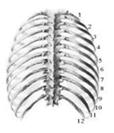
| Fig.1 Model of thorax | Fig.2 X- ray image | Fig.3 The movement of a rib |
In this research, a new method to recover the shape and respiratory motion of the ribs from X-ray images is proposed. Each rib is considered as a curve and its control points are estimated by the cubic B-spline curve iteration algorithm. Figure 1 illustrates the model of thorax which is formed by the twelve pairs of left -right ribs connected with the spine (backbone). Figure 2 shows an example of an X-ray image of a male subject. The position of each rib will change due to the breathing. Figure 3 presents the movement of rib 2 between two images. Each rib touches the backbone in two places, which are the rib head (caput costae) and process transverses, and each rib rotates centering on the axis which passes these places.
To recover the 3D motion and the control points of the ribs using the 2D control points, the factor decomposition algorithm for projective structure and motion has been developed. The measurement matrix has been built with the corresponding control points of each curve. By means of the control points, instead of all points of a curve, data capacity and computational times are reduced. Furthermore, the precision of matching pairs of a rib in multiple images is improved. The estimation errors are not appeared only due to the 2D control point approximation but also the computation of the 3D control points. The experiment with X-ray images has been performed and the effectiveness of the proposed technique has been confirmed through the results with acceptable errors. This approach can be applied to examine the chest expansion for inspecting the pulmonary diseases.
Publications
- Myint Myint Sein et al., "Recovering the 3D Shape and Respiratory Motion of the Rib using Chest X-Ray Image", International Journal of Medical Imaging Technology, Vol. 20, No. 6, Nov. 2002, pp.694~702.[pdf]
- Myint Myint Sein et al., "Detecting the Respiratory Motion of the Rib from X-Ray Image", Proceeding of International Conference on Computer Application (ICCA), Yangon, Myanmar, 2006. [pdf]

